Foot Conditions & Treatments
Achilles Tendinitis Overview
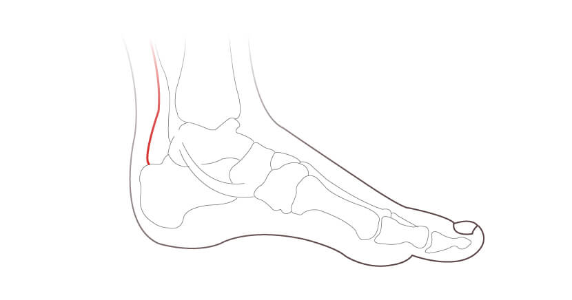
The Achilles tendon is a band of tissue that connects the gastrocnemius and soleus muscle (calf muscle) to the heel bone. Injuries to this tendon are common due to the intense pressure it undergoes during activity. The symptoms that can arise from overuse injuries are broadly known as Achilles Tendinitis.
Achilles Tendinitis Causes
The Achilles tendon is in use during several common activities, including walking, running, jumping and tip toeing. Over time the tendon is worn by age and repetitive activities, making it susceptible to sprains and tears.
Achilles Tendinitis Symptoms and Diagnosis
Symptoms may include:
- Pain and stiffness along the achilles tendon
- Feeling of a lump on the achilles
- Pain at the back of the heel that worsens with activity
- Swelling
- Limited range of motion
The first step to diagnosing this issue would be to arrange an appointment with a podiatric specialist who can assess and advise on treatment. They may refer for an X-ray or MRI.
Achilles Tendon Pain
The pain associated with Achilles Tendinitis often first occurs as a mild ache above the heel or around the back of the leg after activity. More severe periods of pain may then occur after prolonged activity that makes use of the Achilles tendon. The area is often tender or stiff first thing in the morning, although this typically dissipates with movement.
Treatments may include:
Physiotherapy
Your specialist may provide you with stretching exercises that can decrease pain and allow you to return to normal activities.
Orthotics
Your specialist may advise on custom orthotics following a gait analysis. The orthotics can absorb shock to reduce the impact and pain to your heel. A heel raise can also reduce the stress to your achilles tendon as it shortens the distance it needs to stretch during activities.
Shockwave Therapy
Shockwave applies pressure around the affected area with sound waves that pass through the skin to vibrate tissue, which stimulates healing and pain relief. Shockwave therapy is typically carried out once a week for three weeks.
Sodium Hydrochloride and Platelet-Rich Plasma (PRP) injections
When your achilles tendon tries to heal itself, tiny blood vessels can grow from the tendon sheath to the achilles tendon in a region where they are not naturally present. This in turn brings new nerves that can lead to pain. Sodium Hydrochloride (high volume) injections can separate and break off vessels and nerves by gently pushing the tendon sheath away from the tendon. This process helps reset the ineffective healing process and can reduce pain. Following the injection your specialist will advise on an achilles tendon stretching programme for the next two weeks.
Platelet-rich plasma (PRP) therapy uses your body’s own blood plasma with concentrated platelets. The platelets contain growth factors that repair and regenerate damaged tissue. By injecting your own platelets into the affected area the platelets will promote faster healing to the region.
Surgical Management
If symptoms persist then surgery may be suggested. Please click below to find out more on common surgical options.

Ankle arthritis overview
Ankle arthritis is a condition that affects the bones around the ankle joint when the cartilage becomes worn or damaged. Cartilage allows the bones to move against each other without excessive friction and when it becomes damaged the bones rub together causing pain, stiffness and swelling.
Foot and ankle arthritis types
Broadly there are three types of Arthritis of the ankle:
- Osteoarthritis: predominantly occurs in people over the age of 50 and is caused by normal wear and tear of the cartilage over time.
- Rheumatoid Arthritis: is an autoimmune disorder where the body’s immune system attacks its own tissues, particularly the soft tissue in the joints, known as synovium.
- Post-traumatic arthritis: results from an injury that causes premature deterioration of the joint. This may take a long time to develop and the injury may be quite old before the arthritis occurs.
Ankle arthritis symptoms and diagnosis
Symptoms typically include:
- Joint tenderness
- Pain on movement
- Difficulty when moving or putting weight on the foot
- Stiffness, swelling and a warm feeling
- Pain and swelling after rest
Rheumatoid arthritis often occurs symmetrically, meaning that usually both ankle joints are affected at the same time.
Ankle arthritis treatment and prevention
Depending on the underlying cause of the arthritis there are several treatments that could be recommended, including:
- Pain relief, such as NSAID’s
- Pads, arch supports or shoe inserts to stabilise your foot (orthotics)
- Physiotherapy
- Steroid injections
Lifestyle Changes for Foot Arthritis
Many symptoms can be alleviated by changes that you can make to your life style or daily routines. This might include avoiding certain types of exercise, using warm or cold compresses or weight loss. Your specialist will be able to advise you on strategies that could improve your symptoms.
Ankle Arthritis Surgery
Should conservative treatments fail to improve symptoms, surgery may be the recommended treatment.

Acute and chronic foot and ankle injuries are common amongst people of all age groups and activity levels. It is particularly common in people who are active in sports, whether it be recreational or professional. We specialise in accurate diagnosis of your injury with the help of our podiatric, physio and radiology team.
An indication of a foot or ankle issue can include:
- Sharp / Stabbing pain
- Deep aching ankle feeling chronically bruised
- Popping
- Instability
- Swelling with pain
- Locking of the joint
Foot and ankle injuries are typically managed conservatively where you should rest, ice, compress and elevate (RICE). However, further treatment can be suggested, which is why it is important to see a podiatric specialist to review and manage your treatment pathway.
Following your assessment it is likely that an X-ray or MRI will be required to assess the degree of damage.
Treatments may include:
Immobilisation
In order to promote healing and avoid further treatment / intervention, an air cast boot may be provided to keep the area immobile during activity.
Physiotherapy and strengthening
Once adequate healing has taken place our podiatric team may provide you with strengthening exercises. This again can help promote healing and reduce the risk of a repeat injury.
Orthotics, Bracing & Taping
Following an injury, your foot / ankle may become unstable. Your podiatric specialist may advise on custom orthotics following a gait analysis. The orthotics can help improve foot stability and reduce the risk of a repeated injury.
Steroid and Platelet-rich plasma (PRP) injections
If pain persists in your foot / ankle then your specialist may advise on either a steroid or PRP injection.
Typically the choice of substance for a steroid injection will be Cortisone, the steroid can reduce swelling and pain in your foot / ankle. It acts like a hormone, which is made naturally in your body and stops inflammation.
Platelet-rich plasma (PRP) therapy uses your body’s own blood plasma with concentrated platelets. The platelets contain growth factors that repair and regenerate damaged tissue. By injecting your own platelets into the affected area the platelets will promote faster healing to the region.
Surgical Management
If symptoms persist, or you have a repeated or severe injury, then surgery may be suggested. Please click here to find out more on common surgical options.

What is a Bunion?
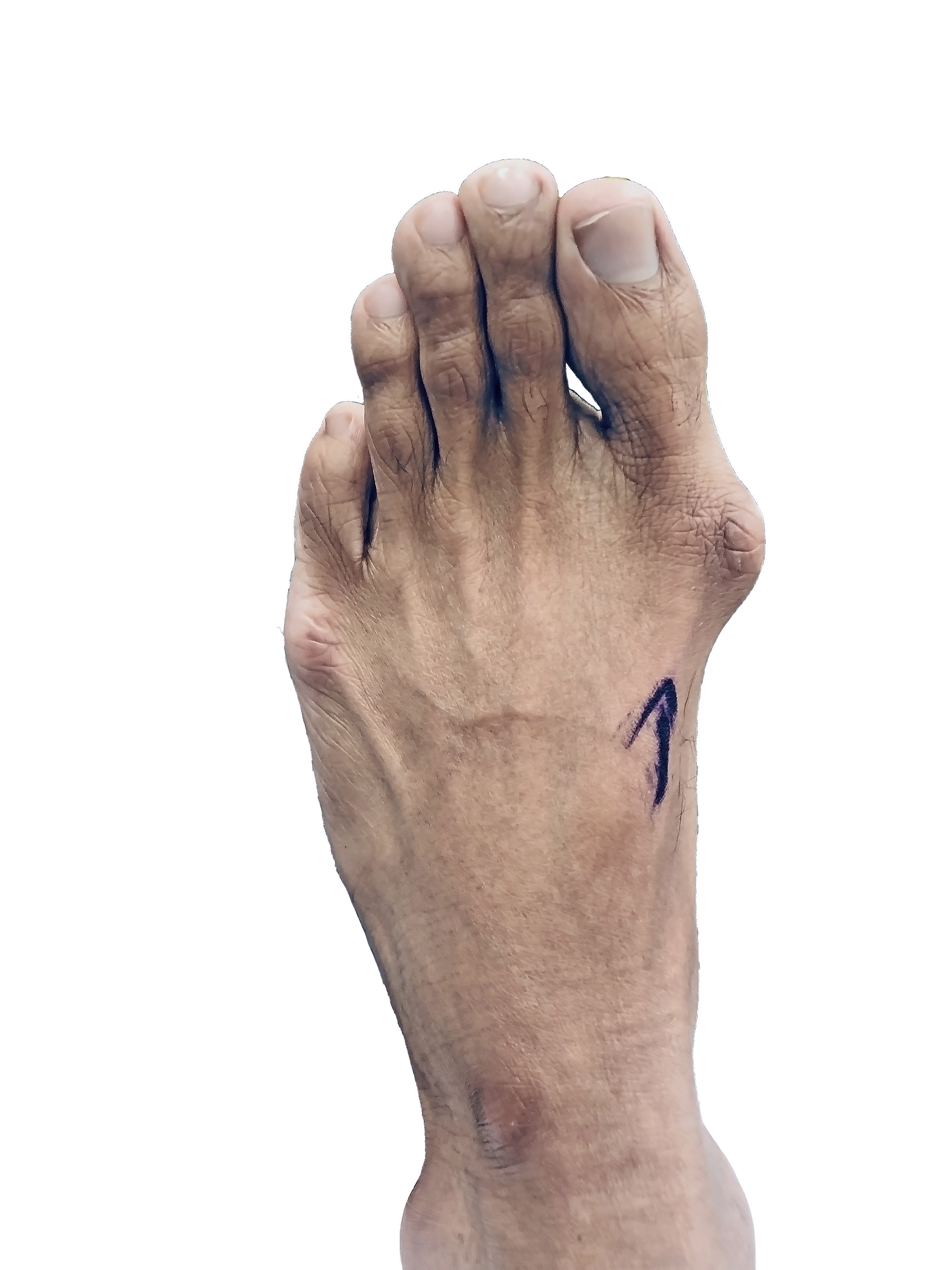
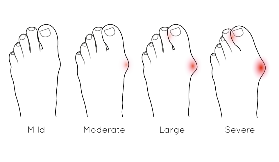
A Bunion, also known as Hallux Valgus, is a common foot condition involving the development of a bony growth below the base of the big toe.
Bunion Symptoms and Diagnosis
A Bunion is a progressive condition, meaning that without intervention it will get worse over time. As the Bunion ‘bump’ grows, the deformity can become increasingly painful when walking or running, cause footwear fitting issues and develop redness and swelling from excessive rubbing in footwear. The position of the big toe will also shift over time, often overlapping the lesser toes, which can cause further pain and discomfort. These symptoms can become quite severe, affecting daily activities and having a significant impact on quality of life.
Causes of Bunions
There is no exact set of causes for Bunions, although likely causes are:
- Hereditary factors – if your parents have Bunions it increases the risk of developing them
- Foot Stress or Injuries
- Arthritis – particularly rheumatoid arthritis
Some experts believe the tight fitting high-pressure footwear, such as high heels, can cause or speed up the progression of Bunions. Certainly they can exacerbate painful symptoms.
Bunion Treatment and Prevention
There are several non surgical treatments for Bunions, including:
- Wearing appropriate shoes with enough space in the toe box
- Using protective cushioning or pads to reduce pain in footwear
- Using toe spacers or night-splints to maintain space between the big toe and lesser toes
- Using orthotics to reduce pressure away from the Bunion
The above measures can help treat symptoms and prevent progression of the deformity. However, currently the only way to fully resolve the Bunion is with surgery. Find out more by clicking the below:

Calf Pain Overview
The calf is made up of two muscles, the gastrocnemius and soleus, which meet at the achilles tendon. Pain in this region is typically related to cramp or sprains.
Calf Pain Symptoms and Diagnosis
If you have strained the calf muscles, symptoms might include:
- Tightness in the calf
- Feeling of a pop, snap or tear at the time of injury
- Sudden pain in the calf
- Difficulty rising onto your toes
- Bruising to your calf
What Causes Calf Pain?
Usually calf pain is caused by muscle injury, though it may also arise from knee problems, foot and ankle problems or nerve problems.
Calf Pain Treatment and Prevention
If you are experiencing calf pain then you can try resting, icing, compressing and elevating (RICE). If symptoms persist then contact a specialist who can assess and advise on a treatment plan.
Treatments may include:
Rest and Immobilisation
Symptoms often result from a tear to some of the muscle fibres and this often requires a period of rest and immobilisation, allowing the muscle to heal. The time required and type of immobilisation depends on the extent of the injury and this will be determined by ultrasound or MRI examination.
Physiotherapy and Stretching
Your specialist may provide you with physiotherapy exercises to stretch the calf muscles, these can relax your calf muscles and in turn reduce the strain it can have to your musculoskeletal system, which might otherwise cause adverse secondary effects such as knee pain and back pain.
Orthotics
Following a gait analysis, your specialist may advise on custom orthotics. A heel raise can for example reduce the tension to your calf muscles by reducing the distance they need to stretch.
Steroid and Platelet-Rich Plasma (PRP) injections
If pain persists in your foot / ankle then your specialist may advise on either a steroid or PRP injection.
Typically the choice of substance for a steroid injection will be Cortisone, the steroid can reduce swelling and pain in your foot / ankle. It acts like a hormone, which stops inflammation.
Platelet-rich plasma (PRP) therapy uses your body’s own blood plasma with concentrated platelets. The platelets contain growth factors that repair and regenerate damaged tissue. By injecting your own platelets into the affected area the platelets will promote faster healing to the region.
Deep Vein Thrombosis Warning Signs
It is important to see a specialist if you experience these symptoms as the cause could instead be related to Deep Vein Thrombosis (DVT), where a blood clot can form in a deep vein.
Symptoms related to DVT can include:
- Pain in your leg, typically starting in your calf
- Red or discolored skin on the affected leg
- A feeling of warmth in the affected leg
Call 999 immediately if you experience the following symptoms also, as this may be caused by a pulmonary embolism:
- Shortness of breath
- Chest pain or discomfort that worsens when you take a deep breath
- Feeling lightheaded or faint
- Rapid pulse
- Coughing up blood
Surgical Management
If symptoms persist then your specialist may advise on surgery. Click here to find out more on common surgical treatments.

Corns and Callus Overview
Corns and calluses are hardened layers of skin that form as a result of friction or pressure, typically against shoes.
Difference Between Corns and Callus
Corns are usually smaller in size than calluses. They tend to form at the top of your toes due to friction against footwear. They can be painful when pressed.
Calluses normally form at the sole of your foot, particularly around the heel and the balls of your feet. They normally aren’t painful to the touch.
Corns and Callus Treatment
Wearing shoes with a large toe box area can help reduce the risk of developing corns. It’s important to regularly clean the build up of dry skin by soaking your feet in warm water for 10 minutes and filing with a pumice stone.
If you are diabetic take extra care when filling and ensure that you check for cuts and blisters to avoid the risk of an infection. You may prefer to have regular treatment with a podiatric specialist.
If you find that the corns and calluses still persist then you can arrange an appointment with a specialist who can assist with treatment, which may include:
Debridement
The corn and / or callus will be carefully removed by a podiatrist with the use of a scalpel. Moisturiser may be used to soften the area.
Orthotics
Your specialist may recommend custom orthotics to reduce the friction between footwear and your feet. If you have additional issues, such as bunions and hammertoes, these can cause formation of corns and calluses, orthotics can help by redistributing the weight and supporting the toes affected by bunions and hammertoes.
Do Corns and Calluses Go Away?
Most corn and callus do go away over time if you remove the friction or pressure that is causing them. However, if you are not sure of the cause, or you are diabetic or the corns and callus are very painful, you should seek a specialist opinion.
How do you get rid of Corns and Calluses Permanently?
Once resolved, corns and callus can be prevented by accurate identification and avoidance of the friction or pressure that is causing it.
What Happens if a Corn is Left Untreated?
Corns can lead to more serious issues if left untreated, including:
- Septic Arthritis or Osteomyelitis – a corn infection can travel to the bloodstream, which would require antibiotics.
- Bursitis – a fluid filled sac can develop beneath the skin, which may require treatment with antibiotics, drain the area or inject a steroid.
- Pain – you may unintentionally change your posture to avoid pain, which can cause painful symptoms in the feet, knees, hips and back.
Can a Corn be Surgically Removed?
The short answer is Yes. If you have a persistent corn then your specialist may recommend surgical removal.

Drop Foot Overview
Drop foot is when it is difficult to lift the front of the foot, this can create difficulty when walking, where your foot will drag along the floor. This may cause you to lift your foot higher to compensate and avoid sliding along the floor.
Due to the nature of foot drop it will change your gait, which can cause you to slap your foot down with each step and create a numb feeling to your toes.
Drop Foot Symptoms and Diagnosis
There are several signs and symptoms of Drop Foot that can help diagnose this issue, including:
- Weakness of the foot muscles can lead to frequent tripping and falls
- The foot may be limp and flop to the side of the body – this can often make it difficult to climb stairs
- The foot may be numb
- Drop Foot often affects only one side, especially when caused by a pinched nerve in the back or leg
To prevent the toes from hitting the ground or tripping you may adapt your gait, typically this will be either: - High Gait – where the leg is raised exaggeratedly to clear the foot of the ground
- Semi-circle Gait – where the leg is kept straight but swings to the side in a semicircle
Drop Foot Causes
Foot Drop is caused by weakness or paralysis of the muscles that lift the foot. These may be caused by:
- Nerve injury where a nerve in your leg that controls the muscles and lift your foot are compressed
- Nerve injury where you have a pinched nerve in your spine.
- Inherited muscular dystrophy
- Brain and spinal cord disorders
If you find that your foot is dragging along the floor whilst walking then consult with your specialist. An MRI may be carried out to assess the underlying cause.
Drop Foot Treatment and Prevention
Ankle-foot brace or splint
Following a gait analysis and possible MRI your specialist may suggest an ankle foot orthosis (AFO). The custom brace helps control the position and motion of your ankle, as well as support weakened limbs.
Surgical Options
To find out more on surgical options to assist with flat foot caused by muscular dystrophy please click here. If the underlying cause is due to a nerve injury or brain/ spinal disorder then a neurologist or neurosurgeon may be suggested.

Flat Foot is where the arch of your foot is collapsed, this is also known as a pes planus foot type. It is common in children as they may still be developing their arches. Flat foot can also be broadly characterised by flexible and rigid flat foot deformity. Not all flat feet are problematic or considered pathological.
The arch can also drop when you are older due to weakening of tendons.
Factors that may cause flat foot include:
- An injury to your foot and/or ankle
- Rheumatoid arthritis
- Genetics
- Hypermobility
- Obesity
- Failure of tendons that support the arch
- Tightness of calves
Flat feet can have adverse effects on the body, the issue may cause pain in your feet, ankle, legs, back, knees and hip.
It’s important to arrange an appointment with a specialist who can assess and provide you with a treatment plan.
Treatment may include:
Physiotherapy and Stretching
Your specialist may advise on stretching exercises that can help correct the fallen arch and decrease pain.
Custom Orthotics
Following a gait analysis, your specialist may suggest custom orthotics that can support your arch. The gait analysis will also assess issues that may be causing pain to your musculoskeletal system, such as calf pain and back pain, the orthotics can for example include a heel raise that can reduce the strain to your calf which may be caused by your flat foot issue.
In more severe cases of flat feet your specialist may instead advise on a brace.
Surgical Management
If symptoms persist then your specialist may advise on surgery. Please click here to find out more.

What is Hallux Rigidus?
Hallux Rigidus essentially involves stiffening or locking up of the big toe joint due to an ongoing arthritic process in the joint. The joint cartilage wears down resulting in pain in the big toe joint, stiffness and limited movement.
It affects both men and women equally but can be exacerbated by previous trauma. Often patients present with a stiff big toe or Hallux Rigidus deformity and pain within the big toe joint. An injury to the joint may predispose a person to Hallux Rigidus. It is not unusual for patients to have stubbed the toe, or someone else may have stood on their big toe many years ago. Initially, a patient may have had some temporary pain they did not think much of at that time. However, over time the joint becomes stiffer and painful.
Hallux Rigidus Symptoms
- Pain and stiffness: These are common symptoms of Hallux Rigidus. Pain and stiffness can be more prevalent during activities such as walking, standing or pushing off while running as they engage the big toe joint. The stiffness in the big toe joint can limit range of motion.
- Limited Range of Motion: The range of motion of the big toe becomes limited, particularly during dorsiflexion (bending the toe upward).
- Swelling: Inflammation and swelling may occur around the affected joint, contributing to pain and discomfort.
- Difficulty Walking: As the condition progresses, activities that involve bending the big toe become more challenging, this may alter your gait to compensate for the pain and stiffness.
- Bone spurs: Over time, the MTP joint in the big toe may develop a bony prominence or enlargement, known as an osteophyte. This can also contribute to a visible deformity of the first MTP joint.
Hallux Rigidus Diagnosis:
- Physical examination: Your specialist will examine your foot and the big toe. They will look for signs of swelling, redness or deformity. The range of motion in the big toe joint will be assessed for pain and stiffness.
- Medical history: Your foot and ankle specialist will request information about your symptoms, such as location and triggers of the pain.
- X-rays: An X-ray is commonly used to assess the bones and joints. It is important to assess a big joint deformity with the use of an X-ray to confirm whether there are osteophytes present, or any other abnormalities.
- MRI or CT scan: A magnetic resonance imaging (MRI) or computed tomography (CT) may be suggested in order to provide a more detailed imaging assessment.
- Gait analysis: An assessment of how you walk, called a gait analysis, may be carried out to ascertain how Hallux Rigidus is affecting your biomechanics.
- Arthrocentesis: Where the joint is aspirated using a syringe or needle to remove fluid. The fluid can be analysed by a pathologist to confirm presence of inflammatory cells, crystals (such as in gout or pseudogout), infection and other abnormalities.
Surgical Management of Hallux Rigidus.
- Cheilectomy:
- Arthrodesis (Fusion of the big toe joint):
- Implant arthroplasty:
- Osteotomy:
This procedure can be performed using minimally invasive surgery or a small open incision. This is usually reserved for mild to moderate osteoarthritis of the big toe or Hallux Rigidus, where only the bony enlargement over the top of the first metatarsal causes pain in shoes and impingement, and restricts joint extension. The procedure is very simple in that it can be performed via keyhole method through a small 3 to 4 mm incision made on top of the toe. A small bur is used to remove the bone spur and a camera may be inserted to the joint to ensure that everything is removed.
The patients will need to rest for 48 hours and then they can start walking again. Only a small plaster dressing on the wound will be present after the first 48 hours and they can transition into trainers. Typically, you may not be able to go back to sports for three to four weeks but normal walking will be allowed after the first 48 hours.
This is the gold standard in advanced or significant osteoarthritis of the big toe joint, where most of the motion has already been lost in the big toe joint and there is very little viable cartilage. Patients who are suitable for the fusion procedure often do not walk properly through the big toe joint and walk only using the outside of the foot, resulting in pain in the hips or the knees, this is known as low gear walking.
A fusion procedure is very successful in that it improves the loading of the foot so that you can walk through the stiff big toe joint pain-free. This allows normal gait and studies have shown that this in the long-term prevents other problems that could be associated with the painful big toe joint.
Typically, a 5-6 cm incision is placed on top of the toe and a screw or a plate is used to fuse the joint. Two weeks of complete rest with no more than 10 minutes an hour followed by using a boot or increasing activity carefully in the shoe to 15 to 20 minutes for the next four weeks until bone healing happens at six weeks. Sports may resume after approximately six to eight weeks gently and running may take a little longer.
This is essentially a big toe joint replacement. It is suitable for the elderly patients who require very little ambulation, no long distance walking or hiking. It is not suitable for younger patients or patients with high activity demands, such as sports. Those patients are better suited for a fusion procedure. However, this is a straightforward procedure that allows quick recovery and return to shoes at approximately two weeks, once the swelling has settled. You can walk on the operated foot after the first 72 hours.
Joint implants for big toe joint have not proven successful in very active and young patient groups. The multi-dimensional demands of the joint are different to other larger joints and therefore they tend to fail in high demand patients.
Osteotomy of the big toe involves realigning of the metatarsal bone or the big toe to improve the range of motion in the joint. This is a rare procedure used where healthy cartilage is present but joint position does not allow range of motion in the big toe joint. This is the least used procedure and works well when there is very little, if any, cartilage damage.
Frequently Asked Questions
Hallux Rigidus has been reported with the prevalence of 2.5% of the population, which means that 1 in 40 people will develop osteoarthritis of the big toe joint through their lifetime. It presents equally in men and women and can be related to trauma or genetics.
Hallux Rigidus surgery involves either shaving down of the bone which is known as a cheilectomy procedure or fusion of the big toe joint which stops any movement and therefore results pain in the big toe joint. These are the most common procedures followed by a possible joint replacement or realignment of bones.
The risk factors for developing Hallux Rigidus can include mechanical problems such as flatfoot or short or long first metatarsal bone. Further genetics of developing osteoarthritis due to early wear and tear or previous history or trauma can exacerbate the problem. Wearing inappropriate or tight footwear can also result in Hallux Rigidus.
Treatment for Hallux Rigidus depends on the severity of the symptoms and duration of the problem. In the first stages you may be able to use a rigid-soled shoe or an orthotic insole device to try to protect the movement in the joint. Should that not settle then an injection into the joint of either hyaluronic acid or steroid can help alleviate pain in the joint at least in the short-term but if conservative care fails then surgery is the only way of treating Hallux Rigidus and it is well known that fusion procedure has been present for more than 40 years with high success rates in approximately 97% of patients.
Recovery from Hallux Rigidus surgery depends on the type of procedure. Most procedures will involve a period of two weeks of rest and thencareful mobilisation until bone healing occurs at six to eight weeks. However, the minimally invasive cheilectomy procedure does allow an initial recovery period of 48 to 72 hours where you are resting and then returning back to most activities except for sports which may take three to four weeks.
Hallux Rigidus is an irreversible process in that the cartilage is worn down over time and this cannot be regenerated. The progression of the disease or correction or replacement of the joint problem can be achieved by surgery.

Hammertoe, Claw Toe and Mallet Toe Introduction
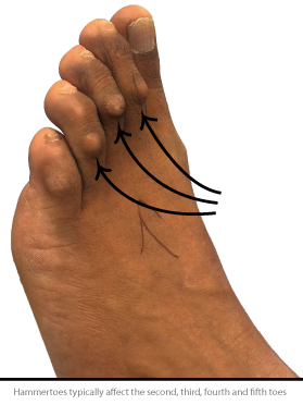
Hammertoe, mallet toe and claw toe describe an issue that develops where the toes are bent. This can create discomfort and friction against footwear, which can lead to additional issues, such as corns, they often coexist with bunions and can often be caused by them.
The difference between these are:
Hammertoe – The toe bends down towards the floor from the middle joint, this then causes the middle toe joint to rise up. This most commonly affects the second toe, but can affect the other lesser toes also. It is less common in the fifth toe.
Claw toe – The toe bends up where the toes and the foot meet and bend down at the middle joint. This often affects all four of the lesser toes.
Mallet toe – The toe bends down at the joint closest to the end of the toe. It most commonly affects the second and third toes but can affect other lesser toes.
Hammertoe, Claw Toe and Mallet Toe Symptoms
The toe issues can create pressure on the foot when wearing shoes and cause discomfort when walking. The joints themselves can be arthritic and painful.
Hammertoe, Claw Toe and Mallet Toe Causes
Toe issues occur due to an imbalance in the muscles, tendons or ligaments that normally hold the toe straight. These may be the result of another problem, such as Bunions.
The type of shoe that you wear, your foot type or injury can exacerbate and contribute to the development of these issues.
Hammertoe, Claw Toe and Mallet Toe Treatment
Unless the problem has progressed to a stage that requires toe surgery, simple conservative measures are recommended in the first instance. These might include:
- Anti-inflammatory Medicines: To relieve pain and decrease inflammation
- Injections: A cortisone injection can further help relieve pain and inflammation in more severe cases
- Physical Therapy: Physiotherapy can be useful to stretch tight muscles and tendons that are causing the hammertoe
- Bespoke Orthotics: A biomechanical specialist can design and manufacture a custom insole to resolve discomfort and prevent progression of the deformity
- Toe Splints or Pads: Splints and pads can be helpful to realign the affected toe and prevent discomfort when walking in footwear
Toe Surgery
If more conservative measures are not successful in eradicating symptoms then surgery may become the recommended treatment.

Our central London clinic is home to consultants who specialise in treating heel pain. There are many causes of heel pain and a range of diagnostic tools are used to evaluate the condition and recommend the most appropriate treatment plan.
Plantar fasciitis is the most common cause of heel pain. This condition typically manifests as pain first thing in the morning when you get out of bed or from resting on a chair. The most common area of pain is on the inner side of the heel and can be described as sharp, burning and throbbing in nature. It typically gets better after the morning and returns towards the end of the day.
Causes of Heel Pain:
- Nerve impingment
- Calcaneal Fracture
- High Arched Feet
- Tight Calves
- Plantar Fascia Tear
- Heel Bursitis
- Plantar Fasciitis
- Back Problems
We use X-rays, Ultrasound and MRI scans as required to ensure that a correct diagnosis of the heel pain and underlying conditions is achieved before any treatment is initiated.
Heel Pain Treatment:
Shockwave Therapy
This non-invasive technology allows for the fascia to heal by encouraging increased blood flow and healing factors from the body to be activated. It has a growing evidence base in treating plantar fasciitis, as well as achilles tendonitis.
Typically three sessions are required, once per week over a three week period. At the final session your specialist will advise whether further treatment is recommended.
Orthotics/Insoles
Custom orthotics can improve foot function, cushion the heels and reduce arch strain. They are effective for most patients suffering from heel pain. A gait analysis and biomechanical assessment will be carried out prior to prescribing custom orthotics. A 3D scan of your feet will then be taken to accurately measure to contours of your feet.
Injections
In recurrent cases, we may recommend an injection of cortisone or plasma rich protein (PRP). These are effective in reducing heel pain and are performed accurately by using ultrasound guidance.
Physiotherapy
Physiotherapy is used in combination with all the above treatments to ensure maximal effectiveness and rehabilitation. In some cases our team will provide exercises that can be beneficial for heel pain, we may otherwise refer to a physiotherapist.
Surgery
Less than 1% of cases require surgery and it is reserved as a last resort. Click below for find out more.
Schedule a consultation with our specialists to assess and discuss options.
Our podiatrist, Mr Steven Thomas, offers a range of non invasive treatments for heel pain. Mr Thomas is covered by most major insurance companies, excluding AXA PPP and Bupa.
Are you covered by AXA PPP or Bupa? Arrange your consultation with Mr Kaser Nazir who is covered by all major insurance companies.
Are you interested in assessing your gait analysis and biomechanics?
Book online today for your gait analysis with our podiatrists at 17 Harley Street.
Self-funding fees for common heel pain investigation and treatments
- Consultation with Mr Steven Thomas: £150
- Gait analysis with Mr Steven Thomas: £220
- Consultation and orthotics package with Mr Steven Thomas: £500
- Shockwave therapy per session: £210

Ingrown toenails develop when the sides of the nail cut and pierce into the surrounding skin. This can cause pain, redness, swelling and possibly an infection.
Common causes of an ingrown can include damage to the nail following a sports injury, an object dropping on the nail or alternatively inappropriate cutting of the nail.
Symptoms of Ingrown Toenail
Signs of an infection may include:
- Swelling
- Redness
- Pain
- A hot sensation in the area
- Pus
- Bad odour
If you do suspect an infection, or pain does not settle after a few days, then arrange an appointment with your specialist as soon as possible.
What causes ingrown toenails?
Causes of ingrown toenails could include:
- Poor nail cutting
- Hygiene
- Predispositions with wider or misshapen toenails, these can be difficult to cut properly.
Diagnosis for Ingrown Toenail
The diagnosis is generally made when you consult a podiatrist or a foot specialist, or alternatively by your GP.
Your specialist may prescribe antibiotics if the area is infected. This may be caused by the nail pushing in to the skin around the nail.
How to get rid of an ingrown toenail overnight?
It’s possible to bathe in salt water and try to remove a small spike of nail, if you are not cutting through the skin and if it’s not painful.
However, if ingrown toenail surgery is required then it is important to see a podiatrist or foot specialist who can assist in taking out the ingrown toenail.
Treatments may include:
Conservative Cut Back
If the issue is not too severe or infected, then your specialist may just need to cut or trim the nail. This can stop the nail from pushing into your skin and subsequently reduce pain, swelling and reduce the risk of an infection. A procedure may be advised at a later stage, especially if the ingrown nail is a recurrent problem.
Antibiotics
If the area is infected then your specialist may provide you with antibiotics, typically this is taken for one week. If symptoms persist following, then a procedure may be suggested.
Surgical Options
If you have a persistent ingrown toenail then your specialist may advise on a nail surgery procedure. Please click here to find out more.
How to prevent ingrown toenails
Prevention is very much dependent on keeping the area clean and cutting appropriately. It is important to wear properly fitting shoes, as this may cause an ingrowing toenail.
When to see doctor or specialist
If there is a sign of an ingrown toenail infection or the ingrown toenail is persistent then you should arrange an appointment to see a specialist. If the ingrown toenail has recurred a podiatrist can discuss surgical treatment options, such as the Zadik’s procedure or an exostectomy procedure if a boney spur is suspected to be the cause.
Ingrown Toenail Surgery: Frequently Asked Questions
Symptoms of ingrown toenail could be from minor pinching in shoes versus local inflammation regularly or a chronic infection where the body is trying to get rid of the ingrown toenail and becomes infected with granulation tissue, which is essentially foreign body reaction.
An ingrown toenail surgery could include partial or full removal of the nail under local anaesthesia. The nail removal surgery may involve a chemical called phenol to help prevent the ingrown toenail from recurring.
Alternatively surgery such as the Zadik’s procedure could be advised. The nail bed is cut out by exposing the nail bed and removing the ingrown toenail. It is then stitched back so that the skin is cut is covered again
Signs of an infected toenail may include pus, swelling, a foul smell, redness of the region and pain
It is important to properly clean and cut the nail to avoid ingrown toenails
- Wearing shoes that crowd the toenails.
- Cutting toenails too short or not straight across.
- Injuring a toenail.
- Having very curved toenails.

Morton’s Neuroma Overview
A Neuroma is a damaged or irritated nerve located between the toes. This can be painful and uncomfortable, which in turn can cause difficulty with footwear.
A Neuroma typically affects the nerve between the 3rd and 4th toes, which is known as a Morton’s Neuroma, and somewhat less often between 2nd and 3rd.
Morton’s Neuroma Symptoms and Diagnosis
Morton’s Neuroma symptoms typically include:
- Sharp radiating pain
- The sense of walking on pebble
- Numb or tingling sensation at the toes
Painful symptoms can worsen over time, especially if you exacerbate symptoms with poor fitting footwear or strenuous activity.
Morton’s Neuroma Causes
There are several causes of the irritated and damaged nerve that causes Morton’s Neuroma. It is often associated with:
- High activity, such as strenuous walking, running or exercise
- High heeled, pointy or shoes that put a lot of pressure on the toes
- Other foot conditions, such as Bunions, Hammertoes or Flat Feet
Morton’s Neuroma Treatment and Prevention
Simple treatments include:
- Wear appropriate shoes that take pressure off the affected area
- Maintain a healthy weight, which can relieve foot pressure and decrease symptoms
- Over the counter pain medication can be used to help control the pain
- Orthotics can help redistribute your weight across the foot and away from the neuroma location
Injection therapy
Your specialist may use steroid or alcohol injections with anesthetic to reduce inflammation and painful symptoms. This resolves a neuroma in around 50% of cases.
Neuroma Surgery
Surgical treatment of neuroma can be used should other treatments fail.

The plantar plates are thickened ligaments found at the balls of feet. An injury or tear to the plantar plate can affect the stability of a toe, this can cause pain and develop issues such as a hammertoe. This most commonly affects the second toes.
There are two forms of plantar plate injury:
Chronic injury – where the injury develops slowly over time caused by micro-tears that stretch the ligament.
Acute injury- where the injury tends to develop following a sudden upwards movement of a toe.
If you experience pain in your toe, ball of the foot or the toe starts to float from the ground then the first step would be to arrange an appointment with your foot specialist.
Treatments may include:
Strapping or a Splint
Your specialist may advise on strapping the toe or provide you with a splint. This will keep your toe aligned and allow the injury to heal.
Immobilisation
Your specialist may advise on immobilisation with the use or a cast or air cast boot and crutches. This again will allow the toe to heal.
Surgical Options
If the injury is not caught soon enough then surgery may be suggested if pain persists. Please click here to find out more.

Brachymetatarsia, is a foot deformity that has the appearance of a shortened toe, this is typically caused by a growth disturbance to the bone. Any toe can be affected by brachymetatarsia but it most commonly occurs to the 4th toe.
It is common to have callus buildup due to the excessive friction against footwear, well fitted shoes can help reduce the buildup.
Treatment may include:
Orthotics
Your specialist may suggest custom orthotics to help redistribute your weight and reduce the risk of your toe rubbing against shoes, this can stop callus build up.
Surgical Management
Surgery may otherwise be suggested to lengthen the toe. Please click here to find out more on common surgical options.

Tailor’s Bunion Overview
A Tailor’s Bunion, or Bunionette, is a prominence of the bone just below the base of the little toe.
A Bunionette can develop over time, where the prominence becomes larger and slowly pushes the little toe in towards the other toes. The underlying cause may well be splaying of the metatarsal bones or outward bowing of the fifth metatarsal bone.
Tailor’s Bunion Symptoms and Diagnosis
The bony growth can cause the metatarsal head to be irritated during activity or in footwear and result in a redness, swelling and pain.
Tailor’s Bunion Causes
The precise cause of a Tailor’s Bunion is not known, although it is linked to:
- An imbalance in the foots structure leading to an outgrowth to the side of the foot. This growth can be exacerbated by pressure and rubbing from footwear.
- A bony spur growing out from the side of the foot
Although it is not thought that tight or heeled shoes cause a Tailor’s Bunion, they do exacerbate painful symptoms and may aid in progression of the issue.
Treatment
A Tailor’s Bunion can be managed with orthotics (shoe inserts) that can cushion the area and redistribute pressure to relieve symptoms. This treatment is often more effective with bespoke orthotics, which are moulded and designed to your particular foot type.
Wearing appropriate footwear that has plenty of space in the toe boxes is also helpful, alongside over-the-counter pain medication to manage the pain.
A steroid injection in to the bursitis that may develop can be helpful.
Tailor’s Bunionette Surgery
If more conservative measures don’t resolve symptoms then surgery may be a sensible treatment option. Find out more here.

Verrucae Overview
A Verruca, also known as a Plantar Wart, is a lump of tough skin that grows on the sole of the foot. They are recognizable by an area of hardened skin, often with a small black dot beneath the surface.
Verrucae Symptoms and Diagnosis
Verrucae can be painful, especially when walking; the sensation is often compared to walking on a needle.
Verrucae Cause
Verrucae are caused by a virus, known as the Human Papilloma Virus (HPV), which is contagious via direct contact that causes other warts such as genital warts and cold sores.
However, when it affects the foot, it is known as a verruca or a verruca pedis and occasionally also terms used such as plantar wart of the foot.
A verruca can occur when there is a break in the skin surface, allowing the virus to enter and cause an infection. Typically you may contract a verruca or wart if you walk barefoot on a rough surface or in a communal area, such as a public swimming pool.
Verrucae Treatment and Prevention
Verrucae will eventually go away by themselves, as your body naturally fights the virus that causes them. However, this can take a long time, sometimes years. As a result, treatment is often sought to quickly resolve the problem.
A verruca is extremely contagious, wearing a plaster over a verruca, or flip flops rather than being barefoot, can help to prevent the spread of the virus.
Treatment
Verrucae can often be treated with over-the-counter creams and ointments that can be obtained from the pharmacy.
If over-the-counter medicines fail to resolve the problem, then other treatments can be tried, including:
- Cryotherapy – where the verruca are frozen off
- Acid Treatments – where the verruca is burned off with an acid, such as Silver Nitrate
If these kinds of measures fail to eradicate the verruca, then surgical removal may be recommended. Click below to find out more about Verruca Surgery:
Verruca Removal: Frequently Asked Questions
A verruca can take months to a few years to go away on its own without treatment. On average:
- In children, verrucae may resolve within 6 months to 2 years, as their immune systems tend to clear the virus more quickly.
- In adults, they may take 2+ years or even longer to disappear naturally, or they may persist indefinitely if the immune system doesn’t clear the virus effectively.
The risk of scarring following verruca removal treatment depend on which treatment is carried out. There is moderate risk of scarring for cryotherapy treatment for example, especially with repeated treatments or deep freezing. Surgical removal has a higher risk of scarring, especially if the wart is large or deep.
The main cause of a verruca (plantar wart) is the human papillomavirus (HPV)
The best treatment to remove a verruca can vary with each individual, it is important to discuss with a podiatric specialist who can review and advise on options.
Home remedies have been varied and include old mother’s tales, such as banana skin or tea tree oil. It seems that over-the-counter treatments such as salicylic acids are the best way to try to treat a verruca at home.
In most cases, verrucae are not harmful — they’re benign (non-cancerous) skin growths caused by the human papillomavirus (HPV). However, they can be bothersome or problematic in some situations.
